Acute Achilles tendon rupture
BMJ 2015; 351 doi: https://doi.org/10.1136/bmj.h4722 (Published 22 October 2015) Cite this as: BMJ 2015;351:h4722
- Dishan Singh, consultant orthopaedic surgeon
- dishan.singh{at}rnoh.nhs.uk
What you need to know
Patients with an Achilles tendon rupture often report feeling a blow to the heel and sometimes an audible snap while playing sports or running
Patients may still be able to walk on tiptoes and plantarflex against resistance because the other ankle plantarflexors are intact
The Simmonds’ triad of altered angle of declination (the foot of the injured leg rests in a more dorsiflexed position than the other side when the patient lies prone), palpable gap, and lack of plantarflexion on calf squeeze test will detect a rupture in nearly all cases
Imaging is rarely necessary
While playing tennis, a healthy 35 year old man felt as though he had been hit on the back of the lower leg by his opponent’s racquet. He couldn’t keep playing but could walk. He presents the next day with mild bruising, swelling, and weakness while walking. He can walk on tip toes but Simmonds’ triad (calf squeeze, altered angle of declination, and palpable gap) confirms the diagnosis of a ruptured Achilles tendon.
What is an Achilles tendon rupture?
A rupture of the Achilles tendon (fig 1⇓) is a disruption in the conjoined tendon of the gastrocnemius and soleus muscles, usually about 2-6 cm proximal to the tendon insertion into the calcaneus.1 Risk factors include increasing age, Achilles tendonopathy, systemic corticosteroids, previous steroid injections into or around the Achilles tendon, and use of quinolone antibiotics.1 2

Fig 1 On opening the paratenon at surgery, the two ends of a complete transverse rupture of the Achilles tendon, with an intervening gap (arrow), are seen. The plantaris tendon is intact (arrowhead)
How common is acute Achilles tendon rupture?
An acute rupture is commonly seen in squash, tennis, football, running, and other sports that require repetitive abrupt jumping or bursts of sprinting2
Injury may also occur from a fall or missing a step or a deep laceration over the site of the tendon. Spontaneous ruptures during normal walking rarely occur
The annual incidence of acute Achilles tendon rupture is around 2 in 10 000
The incidence is rising steadily, possibly due to sedentary lifestyles and intermittent participation in recreational sport in later life3 4
Why is it missed?
A non-specialist may miss the diagnosis in around 20% of cases for several reasons.5 6 The patient may rationalise the injury as a direct blow, leading the clinician to suspect local bruising rather than a rupture.5 The clinician may regard the injury as trivial because a history on an audible snap may not be elicited, about a third of patients with complete tendon rupture do not mention pain,7 external bruising and swelling may be mild (as the tendon usually ruptures within its paratenon sheath), a gap may not be evident in longitudinal tears (fig 2⇓) or with swelling or bleeding, and other plantarflexors may mask the weakness from a ruptured Achilles tendon (fig 3⇓). Thus, a patient being able to walk, stand on tiptoes, and plantarflex the ankle against resistance can lull the clinician into missing the diagnosis.5
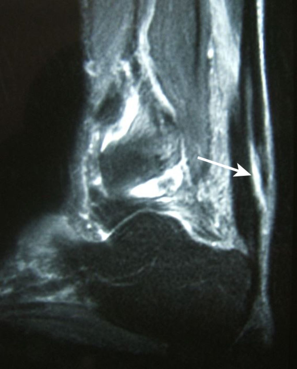
Fig 2 Magnetic resonance imaging of a longitudinal split tear (arrow) in a previously tendonopathic Achilles tendon. No gap in the tendon is evident on palpation

Fig 3 Other tendons (in red) such as the tibialis posterior tendon and the toe flexors can plantarflex the ankle, thus enabling a patient to walk on tiptoes even if the Achilles tendon is ruptured
Why does this matter?
Treatment delay leads to poorer outcomes,1 8 as a discontinuous or lengthened tendon (fig 4⇓) can cause weak plantar flexion, fatigue, limp, and inability to run, heel rise, play sports, and climb stairs.8 More complicated surgery, with longer scars and higher risks of complications, may be required, and return to sports is not always possible.8

Fig 4 Magnetic resonance imaging of a missed rupture at six weeks shows that the two ends of the Achilles tendon have retracted and the gap is filled with a flimsy pseudotendon (arrow), leading to a lengthened and weakened tendon
How is it diagnosed?
Clinical features
Patients with acute ruptures generally describe feeling a sudden painful blow to the heel, sometimes accompanied by an audible snap, during sports or running. They may wrongly believe that they have been kicked or hit on the back of the heel tendon by a ball or racquet. They experience residual calf ache, mild bruising, swelling, and a weakness in “pushing off” with the affected foot.
On examination, ask the patient to lie prone on an examination couch with feet dangling over the edge. Use of Simmonds’ triad (angle of declination, palpation for a gap, and calf squeeze; see fig 5⇓ and video at https://youtu.be/8PvgvUV8N8U),9 yields a more accurate diagnosis than Thomson’s calf squeeze test alone.10 11

Fig 5 Compare both legs with the patient lying prone on a couch and the feet dangling over the edge. External bruising and swelling may be mild, as in this case, and relying on such signs may lead some to miss an acute rupture of the Achilles tendon. However Simmonds’ triad of tests increases diagnostic accuracy. (A) Look for a difference in the angle of declination (injured right foot lies in a more dorsiflexed position). (B) Look or feel for a gap in the tendon (position of finger). (C) Squeeze the calf muscles: plantarflexion of the foot should occur if the tendon is intact (left foot), but may not occur if the tendon is ruptured (right foot)
Simmonds’ triad:
Look (inspect for angle of declination of the foot)—The altered angle of declination (or “angle of dangle”) refers to the loss of tension in a ruptured Achilles tendon, which causes the injured ankle and foot to lie in a more dorsiflexed position than the uninjured one. This test was modified by Matles,12 who asked the patient to actively flex the knee through 90° while observing whether the foot of the injured leg falls into a more dorsiflexed position. The Matles test alone has a sensitivity of 0.88 and a positive predictive value of 0.92.11
Feel (palpate for a gap)—A gap can often be discerned by palpating the tendon along its entire length; the sensitivity is only 0.73 and the positive predictive value is 0.82.11
Move (calf squeeze)—The examiner sequentially gently squeezes the patient’s calf muscles: squeezing the calf deforms the soleus muscle, causing the overlying gastrocnemius-soleus tendon to bow away from the tibia, resulting in plantar flexion of the foot if the tendon is intact.13 If there is an acute rupture, the injured foot remains in its neutral position on squeezing the calf, with a sensitivity of 0.96 and a positive predictive value of 0.98.11
The Simmonds triad is preferred to Thomson’s isolated calf squeeze test,10 as a comparative study found that two of the three tests (palpation, Matles test, and calf squeeze) were positive in all ruptures, suggesting a sensitivity of 100% for this triad.11
Record calf squeeze findings as “plantarflexion noted” or “plantarflexion not noted” (since “calf squeeze test positive” could mean either positive plantarflexion or positive rupture).
Investigations
Imaging is usually unnecessary, as the diagnosis can be made clinically. Ultrasonography or magnetic resonance imaging may help if the diagnosis is unclear (partial rupture or tendonopathy). However, when an acute Achilles rupture is suspected, it is preferable to refer the patient on the same day to an orthopaedic surgeon rather than request investigations that may delay treatment.14
How is it managed ?
Prognosis is good with prompt treatment: normal walking and stair climbing should be possible at a median of 12 weeks after treatment, and return to sports at 9 months.15 The goal of treatment is to restore continuity and normal length and tension of the Achilles tendon. Treatment may be surgical or non-surgical, there is no clear evidence for an optimal method.16 Although surgery ensures a correct apposition of the tendon ends (hence a lower re-rupture rate), complications such as wound breakdown and wound infection may occur. Return to pre-injury level of activity may be achieved without surgery, especially with accelerated functional rehabilitation and early motion.16 17 Non-surgical management is generally best for older, less active patients or those with comorbidities. Surgical management is usually recommended for young people, athletes and people with high levels of activity, and those in whom non-surgical management has been unsuccessful. All patients, whether treated surgically or non-surgically, should have supervised physiotherapy for several months.
The risks of thromboembolic episodes18 after acute Achilles tendon ruptures have been found to be high in some studies: discuss the risks and benefits of thrombo-prophylaxis with the patient and monitor carefully for this complication.
Notes
Cite this as: BMJ 2015;351:h4722
Footnotes
This is one of a series of occasional articles highlighting conditions that may be more common than many doctors realise or may be missed at first presentation. The series advisers are Anthony Harnden, professor of primary care, Department of Primary Care Health Sciences, University of Oxford, and Richard Lehman, general practitioner, Banbury. To suggest a topic for this series, please email us at practice@bmj.com.
Competing interests: I have read and understood the BMJ Group policy on declaration of interests and have no relevant interests to declare.
Provenance and peer review: Not commissioned; externally peer reviewed.